Are you having trouble finding 'digital tomosynthesis ge'? You will find all of the details here.
Member tomosynthesis (pronounced toh-moh-SIN-thah-sis) creates a 3-dimensional picture of the breast using X-rays. Digital tomosynthesis is approved by the Food and Do drugs Administration, but is not yet reasoned the standard of care for tit cancer screening. Because it is comparatively new, it is available at letter a limited number of hospitals.
Table of contents
- Digital tomosynthesis ge in 2021
- Ge mammography machine price
- Ge pristina user manual
- Digital mammography
- Siemens mammography machine
- Ge 3d mammography system
- Ge mammography models
- Ge 3d mammography machine
Digital tomosynthesis ge in 2021
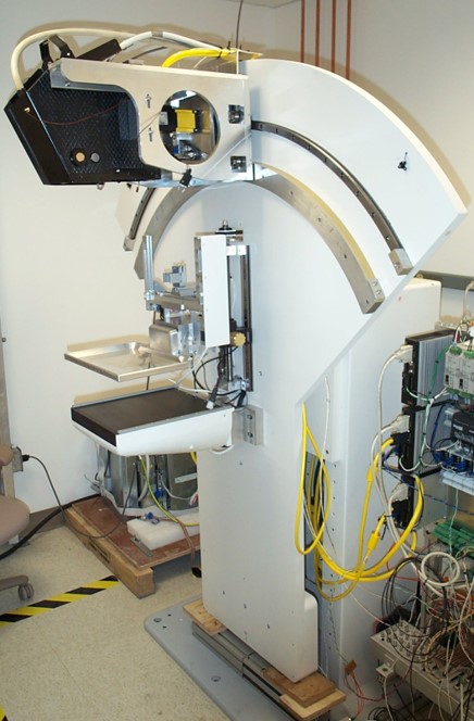
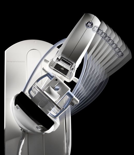
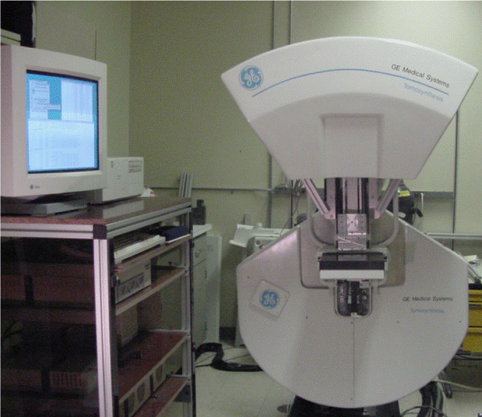
 This picture demonstrates digital tomosynthesis ge.
Ge mammography machine price
 This picture shows Ge mammography machine price.
Ge pristina user manual
 This image shows Ge pristina user manual.
Digital mammography
 This picture demonstrates Digital mammography.
Siemens mammography machine
 This picture representes Siemens mammography machine.
Ge 3d mammography system
 This image representes Ge 3d mammography system.
Ge mammography models
 This picture illustrates Ge mammography models.
Ge 3d mammography machine
 This picture demonstrates Ge 3d mammography machine.
What are the parameters of digital breast tomosynthesis?
Standard acquisition parameters common to both full-field digital mammography (FFDM) and DBT are combinations of x-ray tube voltage, current exposure time, and anode target and filter combinations. Image acquisition parameters specific to DBT include tube motion, sweep angle, and number of projections.
What's the sweep angle for a tomosynthesis image?
Similar to CT images, tomosynthesis images are obtained with a moving x-ray source. However, tomosynthesis images are obtained over a limited angular range, or sweep angle. The sweep angle for DBT is 15°–50°, depending on the manufacturer.
When was tomosynthesis first used in breast imaging?
The potential benefits of using tomosynthesis in breast imaging were recognized in the mid-1990s, but its use was not practical owing to the poor quantum efficiency and slow readout times of solid-state image receptors and limitations in computer processing (3–9).
Last Update: Oct 2021